Clinical-trial imaging analytics

Earlier proof that
neuro-recovery treatments
are working.

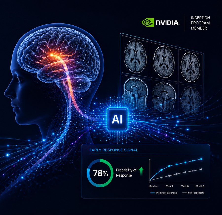
AI analysis of routine MRI scans to generate early treatment-response signals for stroke, traumatic brain injury, and spinal cord injury trials.

Request a pilot See the science
Stroke TBI Spinal cord injury Phase II / III trials Pharma & biotech sponsors
6mo
Typical endpoint wait time
$1M+
Daily Phase II/III trial burn
0
Early response signals available today
The problem

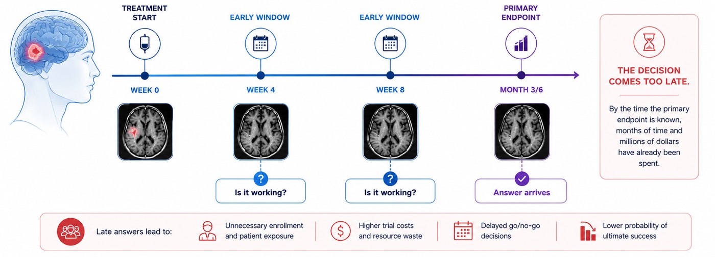
The decision comes too late.

Patients recover at very different rates. Sponsors typically wait months for a clinical endpoint to read out — months of uncertainty that translate directly into wasted enrollment, wasted trial cost, and delayed go or no-go calls.

W0
Treatment start
Trial begins. Baseline MRI collected. Clock starts.
W4
Early window
Is it working? No answer available.
W8
Mid window
Still no signal. Trial spend continues.
M6
Primary endpoint
Answer finally arrives. Too late for early decisions.
The solution

Earlier signal. Better decisions.

Axonomics doesn't replace the trial. It improves the timing and quality of the decision — using the same routine MRI scans the trial already collects.

Without Axonomics
Decision waits for the primary clinical endpoint
High uncertainty during the early window
Continued spend on programs that may not be working
Difficult conversations with board and investors
With Axonomics
Earlier go or no-go support in the trial
Better patient selection in future cohorts
Stronger endpoint and trial-design confidence
Clearer story for board and investors
How it works

MRI scans in. Early response report out.

No new scanners. No new protocols. Fits inside the sponsor's existing imaging workflow. Output: an auditable report, not a research file.

01
Routine MRI collected
Standard imaging already captured during the trial. Existing scanners and protocols.
02
Secure upload & de-identification
Compliant data transfer from sponsor or imaging CRO. Audit trail on every step.
03
AI extracts recovery patterns
Models identify imaging features associated with neurological recovery.
04
Signal vs. expected recovery
Each patient scored against a model of natural recovery for that indication.
05
Sponsor report delivered
An auditable readout to support trial-decision conversations with the team.
Scientific basis

The signal is already in the scan.

Routine MRI contains biological information correlated with neurological recovery. The data exists. What's required is the model to extract it.

1,715
Real-world stroke registry
A contemporary, real-world acute stroke imaging registry with linked NIHSS outcomes. Candidate training cohort for stroke trajectory modelling.
3,000
TRACK-TBI — traumatic brain injury
MRI with GOSE outcome measures. Key dataset for TBI recovery pattern extraction.
4,500
CENTER-TBI — European TBI cohort
Multi-site European dataset with detailed outcome labelling across injury severity.
4
MRI signals that matter
Lesion & edema volume · White matter integrity · Cortical volume · Spinal cord signal change. All from standard sequences already collected.
What a sponsor receives.

A synthetic case, shown to illustrate the report format. Real reports are generated from de-identified patient imaging under a sponsor data-use agreement.

Recovery-trajectory tracking
Synthetic case — for illustration
Case ID
SYN-0417
Indication
Ischemic stroke
Imaging timepoint
Week 4 post-baseline
Expected recovery (registry norm) Tracked signal — this patient
W0 W4 W8 W12
ON TRACK

Tracking above expected recovery trajectory at Week 4.

Model in development. Not for clinical use. Not FDA cleared. Report format shown for illustration; case data is synthetic.
Real numbers, early stage.

Pilot metrics below. Full-cohort test-set validation is underway.

r ≈ 0.60
Current pilot correlation with NIHSS (R² ≈ 0.35)
~150 · ~300
Planned test subjects · scans (T1w + DWI) at full cohort scale
R² + AUC
Reported once full held-out test-set validation completes

Building — early pilot on a small subset. Full-cohort validation, drawn from a contemporary, real-world acute stroke imaging registry with linked functional-outcome data, in progress.

The team

Built by people who know the problem.

Neuroscience expertise, machine learning depth, and clinical research credibility.

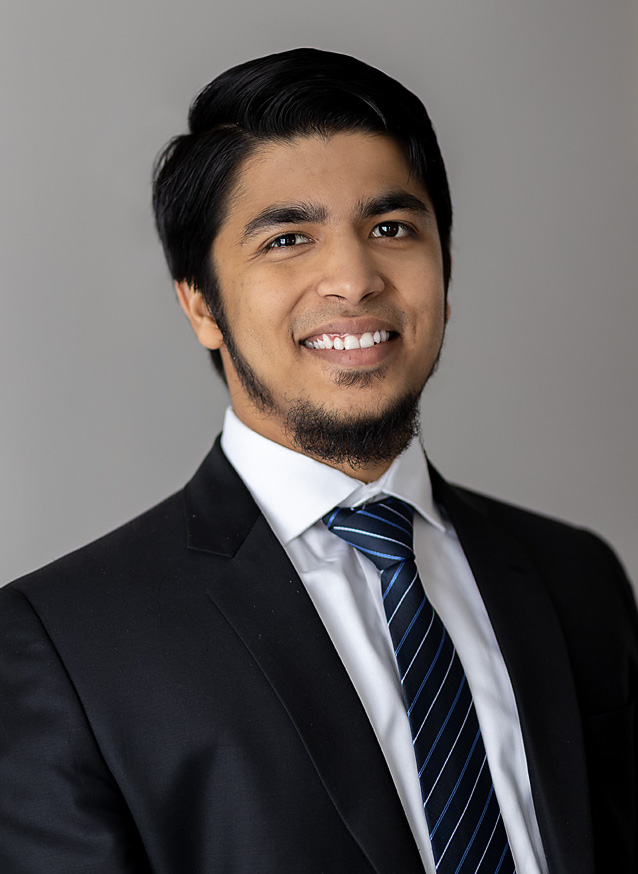
Mohammed Ali Alvi
Mohammed Ali Alvi
Founder & CEO
Dr. Shams Syed
Dr. Shams Syed
CTO & Co-Founder
Michael G. Fehlings
Michael G. Fehlings
Scientific Advisor
Work with us

Run a retrospective pilot
on your completed trial.

Proof-point and revenue. No prospective trial required to get started.

Get in touch How it works